


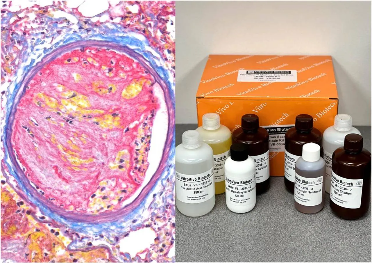
Microscopic tissue review often begins with routine staining, but some research questions require more specific visual information. Methods such as Martius Scarlet Blue MSB stain can help distinguish fibrin and related tissue components when general morphology alone does not provide enough contrast.
Special stains are useful because different biological materials respond to different staining chemistries. Selecting the right method allows researchers to focus on the structure or deposit that matters most to the study rather than relying on a single stain for every tissue question.
Why Greater Tissue Contrast Matters
A tissue section may contain collagen, fibrin, muscle, erythrocytes, protein deposits, and many cellular structures within a very small area. Routine stains can reveal overall architecture, but closely related components may remain difficult to separate confidently.
Targeted histochemical methods improve visualization by emphasizing selected chemical or structural features. This added contrast can make comparison between tissue regions easier and may help researchers connect microscopic changes with other experimental findings.
Detecting Amyloid Deposits More Clearly
Some studies focus on abnormal extracellular protein accumulation rather than connective tissue or fibrin. In these situations, congo red staining amyloid can help researchers visualize amyloid deposits through the characteristic interaction of Congo Red dye with beta-pleated sheet structures.
The method can be applied to formalin-fixed paraffin-embedded tissue and, depending on the protocol, frozen sections. Amyloid-containing areas appear red under routine light microscopy, allowing researchers to assess the location and distribution of deposits within preserved tissue.
Distinguishing Fibrin and Connective Tissue
Fibrin presents a different histological challenge. It may occur alongside collagen, muscle, and erythrocytes, particularly in research involving clot organization, tissue injury, wound repair, vascular changes, or inflammatory processes.
MSB staining provides multiple colors within the same section. Fibrin and muscle typically appear red, collagen appears blue, and erythrocytes appear yellow. This differentiation helps researchers evaluate several tissue components without requiring separate slides for every structure.
Match Preparation With the Staining Method
Successful staining begins before reagents are applied. Fixation time, section thickness, deparaffinization, hydration, slide adhesion, and reagent exposure can all affect how clearly the final stain demonstrates its intended target.
Researchers should use consistent preparation conditions when comparing multiple specimens. Even small technical differences may change color intensity or background appearance, making biological comparisons more difficult if processing conditions are not standardized.
Include Suitable Positive Controls
Positive control tissue helps confirm that a staining procedure is functioning properly. A known fibrin-containing specimen can support MSB quality assessment, while tissue with established amyloid deposits can help evaluate whether Congo Red staining has performed as expected.
Controls should ideally be processed alongside experimental sections. Keeping section thickness, reagent timing, differentiation steps, and imaging conditions consistent makes it easier to determine whether an unexpected result reflects tissue biology or a technical problem.
Look Beyond Color Alone
Color is an important part of histochemical interpretation, but it should not be the only consideration. Researchers should also review tissue location, deposit shape, structural relationships, and whether the staining pattern fits the expected anatomical context.
For example, a deposit near a vessel may carry different research significance from material located within connective tissue or another compartment. Combining staining pattern with morphology can provide a more complete view than simply recording whether a particular color is present.
Standardize Microscopic Imaging
Digital imaging allows researchers to preserve and compare staining results across a study. However, changes in magnification, illumination, exposure, white balance, or image processing can make similar sections appear different.
Using consistent microscope and camera settings reduces this variability. When quantitative or semi-quantitative analysis is planned, researchers should also establish scoring rules, regions of interest, and measurement criteria before analyzing the complete sample set.
Choose Methods Based on the Question
No special stain is universally better than another. The correct method depends on the tissue component that must be visualized and the type of information needed from the specimen.
Fibrin-focused research and amyloid-focused research may both use histological sections, yet they require different chemical approaches. Defining the research endpoint before staining helps laboratories choose suitable preparation methods, controls, and interpretation strategies.
Conclusion
Specialized histochemistry helps researchers examine tissue components that may not be sufficiently clear with routine staining alone. Different staining methods provide different types of contrast, allowing investigators to focus on structures and deposits relevant to specific biological questions.
Reliable results depend on more than staining chemistry. Consistent preparation, appropriate controls, standardized imaging, and careful morphological interpretation all contribute to stronger tissue analysis and more meaningful comparisons across experimental samples.